实验室
LaboratoryMicroscopic CL
Laboratory of Microscopic CL/EDS and Scanning Microscope
Main instrument configuration:
Order number |
Name |
Brand |
Model |
1 |
Cathodoluminescence |
Britain /CITL |
CL8200 MK5-2 |
2 |
Energy spectrometer |
Britain /CITL |
X2072 |
3 |
Polarization microscope |
Germany /Leica |
DM2700P |
4 |
The microscope of scanning synthesis |
Germany /Leica |
DM6M |
Simple descriptions:
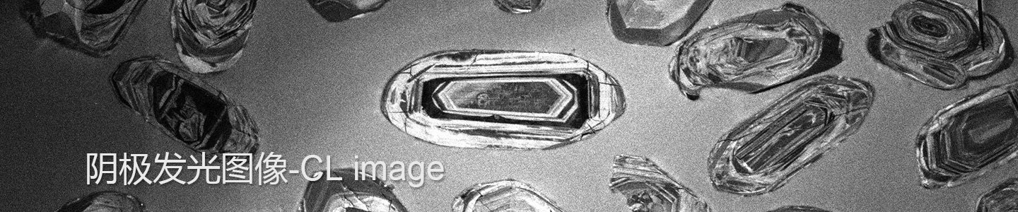
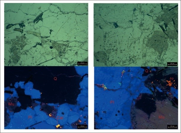
1. Microscopic CL/EDS
The cathodoluminescence and energy spectrometer system of optical microscope is an experimental system that organically combines the mineralogical observation of rock slices under transmission and reflection microscope with cathodoluminescence technology and electronic micro area chemical composition analysis technology. The instrument can realize the conversion of three modes and images of single polarization, orthogonal polarization and cathodoluminescence under the microscope at the same time, and the mineral phase can be semi-quantitatively identified in-situ by energy spectrometer. The use of this instrument can greatly improve the efficiency of the observation of rock slices and mineral identification, and provide a basis for subsequent fine quantitative research.

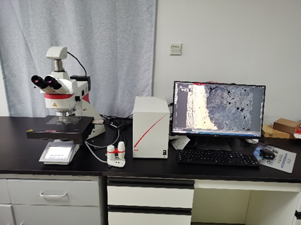
2. Scanning Microscope
The scanning microscope can carry out high-precision morphological analysis on the whole slice. It takes local high-definition photos of each field of microscope, obtains the high-definition microscopic image of each area, and then automatically synthesizes it through software calculation. The instrument can not only obtain the overall image after the synthesis of the whole slice, but also save the high-definition image of each local area. The synthetic image can clearly observe its overall morphology and structure, and also provide great convenience for finding typical minerals. At the same time, the instrument can not only carry out two-dimensional (plane XY axis) automatic scanning, but also carry out more time-consuming three-dimensional (plus Z axis) automatic scanning. Among them, the complete two-dimensional scanning of a slice takes about 25 minutes and the three-dimensional scanning takes about 4 hours.

NOTE: There is a responsible person in the laboratory to assist in the operation.
Welcome to come work with your rock slices!
Responsible people: Hao Deng
Telephone contact: +86-15527381968
Email: denghao815@cug.edu.cn
Location: Room 221, The laboratory building of the SES to the south of the lake, Eastern Campus of China University of Geosciences (Wuhan)